Bio 197 Lab 6 Fungi Learn with flashcards, games, and more — for freeFungi, the microorganisms that grow on everything from plants to people, can be quite eyecatching when viewed under a microscopeUse the space below to draw a picture of the Penicillium specimen as you viewed it under the microscope Basidiomycota (club fungi) View the mushroom specimens available in the lab Do not dissect them See if you can find the gills on the underside of the basidiocarp Name the specific spores formed by the mushroom in the gills

The Many Kinds Of Fungi
Club fungi under microscope
Club fungi under microscope-The fungi comprise a diverse group of organisms that are heterotrophic and typically saprozoic In addition to the wellknown macroscopic fungi (such as mushrooms and molds), many unicellular yeasts and spores of macroscopic fungi are microscopic For this reason, fungi are included within the field of microbiologyObserve a prepared slide of the mushroom Coprinus under the compound microscope A Focus using the 4X lens and locate the edge of a gill Observe with the 10X and 40X lenses B Observe the clubshaped structures (called basidia) on which spores are produced The club fungi derive their name from these clubshaped structures
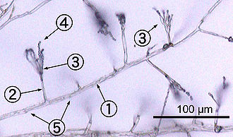


Fungus Wikipedia
Mushrooms under the Microscope Mushrooms, also known as fungi or toadstools, are the sporebearing fruiting body of a fungus The mushroom typically grows above ground or on top of its food source (on a log, or out the side of a mossy tree)The fungi in the Phylum Basidiomycota are easily recognizable under a light microscope by their clubshaped fruiting bodies called basidia (singular, basidium), which are the swollen terminal cells of hyphaeThe basidia, which are the reproductive organs of these fungi, are often contained within the familiar mushroom, commonly seen in fields after rain, on the supermarket shelves, andCharacterized by a basidium Which is a clublike sexual reproductive structure that produces spores
Microscope SlidesTop QualityAffordableBacked by expert technical supportFor over 70 years our mission has been to provide educators with topquality microscope slides for botany, zoology, histology, embryology, parasitology, genetics, and pathology WeFood fungus under the Microscope#FungalELEMENTS#FungusMicroscopy#PenicilliumBasidiomycota The Club Fungi The fungi in the Phylum Basidiomycota are easily recognizable under a light microscope by their clubshaped fruiting bodies called basidia (singular, basidium), which are the swollen terminal cell of a hypha
Mushroom Forceps/Fingers Microscope Slide Dissection Microscope Paper Towels Lab Sheet Light Microscope Put this under the dissection scope (b) Examine them and describe them in the data section) 4) Next, look under the cap and observe the gills (c) Draw what the underside of the cap/gills look like on the data sheet Club Fungi areThe fungi in the Phylum Basidiomycota are easily recognizable under a light microscope by their clubshaped fruiting bodies called basidia (singular, basidium), which are the swollen terminal cell of a hypha Can fungi be seen under a light microscope?Under the microscope you will see the various cells and strands of hyphae that make up the fungus In gilled and poroid fungi basidiomycetes specialised hyphae on the gills known as basidia carry the developing spores and the number of spores on each basidium and the form of attachment vary between species


The Many Kinds Of Fungi
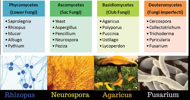


Classification Of Fungi Mycology Microbe Notes
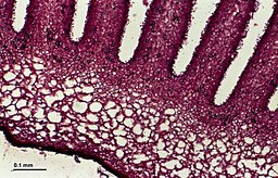
3 Basidiomycota (club fungi) View the mushroom specimens available in the lab Do not dissect them See if you can find the gills on the underside of the basidiocarp Basidiomycota Q1 Name the specific spores formed by the mushroom in the gills Coprinus mushroom View the cross section slide of the Coprinus mushroomThey can be observed under the microscope by taking a scalp but their presence or absence can often be determined in small fruit bodies by simply placing them onto aUnder the microscope, it should now be possible to see any of the important characteristics which will be used for identification These include the spores, acsi (sacs containing the spores), paraphyses (infertile structures between the asci), hairs, and


What Is The Scientific Classification Of Fungi Quora


Experiment 24 General Medical Microbiology Lab
Missouri Mycological Society Welcome to the magical world of mushrooms and one of the most active mushroom clubs out there!Basidiomycota (/ b ə ˌ s ɪ d i oʊ m aɪ ˈ k oʊ t ə /) is one of two large divisions that, together with the Ascomycota, constitute the subkingdom Dikarya (often referred to as the higher fungi) within the kingdom FungiMembers are known as Basidiomycetes More specifically, Basidiomycota includes these groups mushrooms, puffballs, stinkhorns, bracket fungi, other polypores, jellyEssentially, mycology is the study of fungi Here, mycologists directly focus on the taxonomy, genetics, application as well as many other characteristics of this group of organisms Currently, over 50,000 species of fungi have been identified in different environments across the globe While some are freeliving and have no impact on human


2500sched Htm
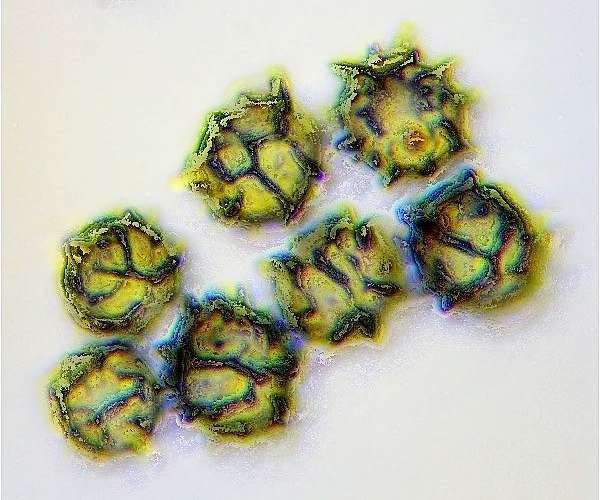


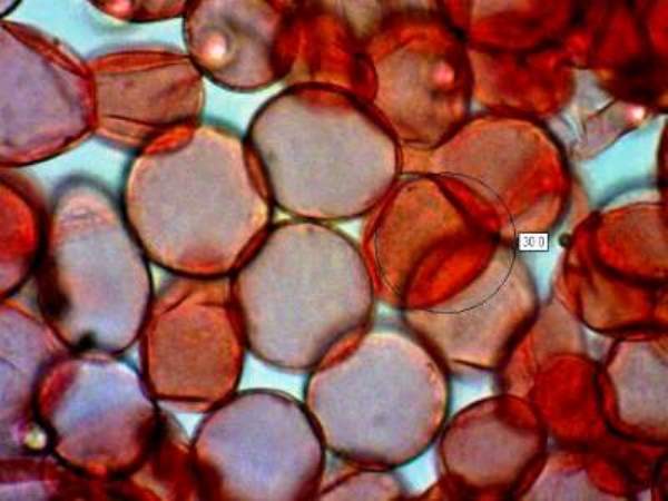
Microscopy Of Spores Hyphae Cystidia Trama To Identify Fungi
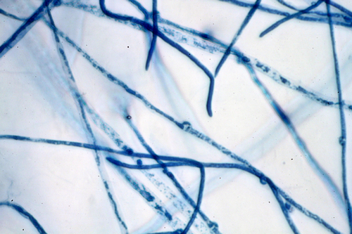
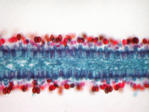
6 Describe what you see in this mushroom section viewed under a microscope and describe where basidiospores are produced in a mushroom, how the structure of the mushroom provides surface area for spore production, and how the spores are dispersed (1 pts) This slide looks like a lake side or sea side rocky edge with a bunch of boats or people swimming to meFungal structures observation under microscopeLactophenol cotton blue (LPCB) PreparationShowingHypaheSepatae hyphaeConidiaCondiophoresSporangium#LPCB#Fungus#The use of a microscope can be fascinating or in some cases frustrating if you have limited experience with microscopy Ideally, if you wish to become proficient at identifying turf diseases, it's best to have a dissecting microscope (640X) and a compound microscope (X) Each microscope is valuable and has particular strengths
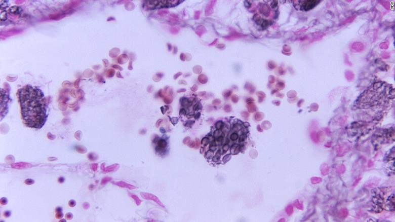


Fungal Diseases Under The Microscope


Club Fungi
The fungi in the Phylum Basidiomycota are easily recognizable under a light microscope by their clubshaped fruiting bodies called basidia (singular, basidium), which are the swollen terminal cell of a hypha This group also includes shelf fungus, which cling to the bark of trees like small shelvesThe Basidiomycota (basidiomycetes) are fungi that have basidia (clubshaped structures) that produce basidiospores (spores produced through budding) within fruiting bodies called basidiocarps ( Figure 532 ) They are important as decomposers and as food This group includes rusts, stinkhorns, puffballs, and mushroomsLichen and fungi under microscope 3D illustration virus bacteria Viral infection causing chronic disease, decreased immunity Red bacteria under microscope closeup Virus abstract background in space Fresh Branching budding yeast cells Colonies of mold fungi cultivated from indoor air


Fungi 1


Microscopy Of Spores Hyphae Cystidia Trama To Identify Fungi
Basidiomycota The Club Fungi The fungi in the Phylum Basidiomycota are easily recognizable under a light microscope by their clubshaped fruiting bodies called basidia (singular, basidium ), which are the swollen terminal cell of a hypha• Another feature of fungi is the presence of chitin in their cell walls • A complex carbohydrate that makes up the cell walls of fungi • The reproductive structure growing from the mycelium in the soil that you recognize as a mushroom • Basilia, a spore making structure in the gills under the fruiting body Characteristics 4Light microscopes are handy optical instruments that come with a variety of essential uses, such as in studying various microorganisms, including parasites, bacteria, and fungi However, they also come with many limitations While a light microscope can be extremely beneficial in bacteriology and pathology, can the same be said for, say


Kingdom Fungi Types Examples Morphology Structure And Importance


3
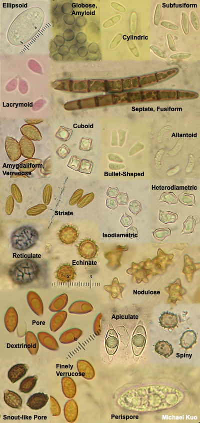
To study fungal spores, basidia, cystidia, sphaerocysts and other tiny features of fungi you will need a microscope capable of at least x 400 magnification Ideally, go for a microscope with a maximum magnification of x 1000, but to obtain reasonably clear images at such high magnification it should have an oil immersion lensUsing your forceps, tear of a small piece of lamellae (gill) from the fungi and mount in water Do this again in Meltzer's ReagentAdd cover slipGently squish the tissue under the cover slip with an eraserFollow these steps when mounting slides onto the microscope 1) Lower microscope stage completely and set objective lens to lowest powerFungal diseases under the microscope Updated 515 AM ET, Tue Share 1 of 9 Tiny fungal spores are found in soil, air and water, and whilst most species are harmless to humans


Fungus Wikipedia


Club Fungi Fungi Painting Club
Come and immerse yourself in the beautiful and mysterious world of fungi If finding, eating, photographing, researching, or just learning about mushrooms sounds interesting, then the Missouri Mycological Society (MOMSGeorgia Regents University (Formerly Augusta State University) Bio 1108 Lab Practical #1 (Protists, Fungi and Microscope) Terms in this set (38) Mechanical Stage club fungiform basidiospores on club shaped structures called basidia mushrooms, puffbals, bracket fungi, rusts, smuts Some images used in this set are licensed under theThis multiplication of yeast cells is usually rapidly occurring, and thus easy to observe on a microscope Of course, the various parts of a yeast cell can also be identified by using a higher powered microscope, or a fluorescent microscope For this purpose, the yeast almost always needs to be dyed in order for the details to become clearly visible


Ascomycota Sac Fungi Basidiomycota Club Fungi Victoria Willis Quyen Mac Ppt Download


Fungi Microbiology
Basidiomycota The Club Fungi The fungi in the Phylum Basidiomycota are easily recognizable under a light microscope by their clubshaped fruiting bodies called basidia (singular, basidium), which are the swollen terminal cells of hyphaeFungi are under the microscope for their medicinal properties Fungi, like mushrooms, have been used for their health properties for millennia Now, mushrooms are increasingly in the public spotlightOur Customer Service team is available from 730am to 8pm, ET, Monday through Friday Live chat is available from 8am to 6pm ET, MondayFriday Call Fax Email


462 Rust Fungi Stock Photos And Images 123rf


Using A Microscope To Study Mushrooms Mushroomexpert Com
You could buy a new microscope, of course You can probably find new microscopes online that meet the requirements above for under $400 at the time of this writing (19) However, and this is a big however, you cannot find a good new microscope for that price With microscopes it is all about the lenses, and there are many knockoffBasidiomycota The Club Fungi The fungi in the Phylum Basidiomycota are easily recognizable under a light microscope by their clubshaped fruiting bodies called basidia (singular, basidium ), which are the swollen terminal cell of a hypha The basidia, which are the reproductive organs of these fungi, are often contained within the familiar mushroom, commonly seen in fields after rain,View our latest service hours »
.jpg)


Superficial Mycoses


Carmarthenshire Fungi December 16
Basidiomycota The Club Fungi The fungi in the Phylum Basidiomycota are easily recognizable under a light microscope by their clubshaped fruiting bodies called basidia (singular, basidium), which are the swollen terminal cell of a hyphaBasidiomycota The Club Fungi The fungi in the Phylum Basidiomycota are easily recognizable under a light microscope by their clubshaped fruiting bodies called basidia (singular, basidium), which are the swollen terminal cell of a hypha The basidia, which are the reproductive organs of these fungi, are often contained within the familiar mushroom, commonly seen in fieldsThe fungi in the Phylum Basidiomycota are easily recognizable under a light microscope by their clubshaped fruiting bodies called basidia (singular, basidium ), which are the swollen terminal cell of a hypha The basidia, which are the reproductive organs of these fungi, are often contained within the familiar mushroom, commonly seen in fields after rain, on the supermarket shelves,


I Barbara Sees A Club Fungus On The Ground And Chegg Com


Club Fungi Microscopic Photography Fungi Biology
Observe the slide under high dry and oil immersion Try to identify the budding cells Make a sketch in the results section Observe the colonies of Aspergillus niger and Penicillium camembertii Describe the morphology and draw an example of each colony in the results section View the plates under the dissecting microscopeMicroscope Bacteria Identification Upon viewing the bacteria under the microscope, you will be able to identify the bacteria based on a wide variety of physical characteristics This mainly involves looking at their shape and size There are a wide variety of different shapes, yet the three main types are cocci, bacilli, and spiralClub fungi Phylum Basidiomycota Include mushrooms, toadstools, puffballs, jelly fungi, shelf fungi, rusts and smuts Larges known club fungus in Oregon is 35 miles across!
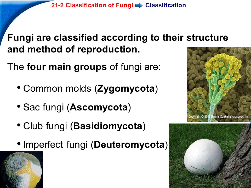


21 2 Classification Of Fungi Ppt Video Online Download


Scanning Electron Microscope Blog April 13
Procedure By using a dissecting needle we prepared a wet mount from the culture provided and made observations under microscope We observed all distinguished featured and recorded by taking a picture Part7 Phylum basidimycota Club Fungi Materials commercial Fungi, demonstration specimen of Club Fungi MushroomsBut,, it takes time, some fungi take months to sporulate The time and the nature of the block of agar are determinant factors This technique allows the intact morphology of the fungus to be seen under the microscope


Basidiomycota


24 3c Ascomycota The Sac Fungi Biology Libretexts


Fungi Protist Prezi By Nini Ho


Fungi Microbiology


Fungi Microbiology


Kingdom Fungi Continued Fungal Phyla 3 Phyla But
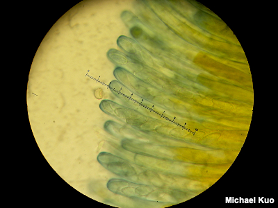


Using A Microscope To Study Mushrooms Mushroomexpert Com


Club Fungi
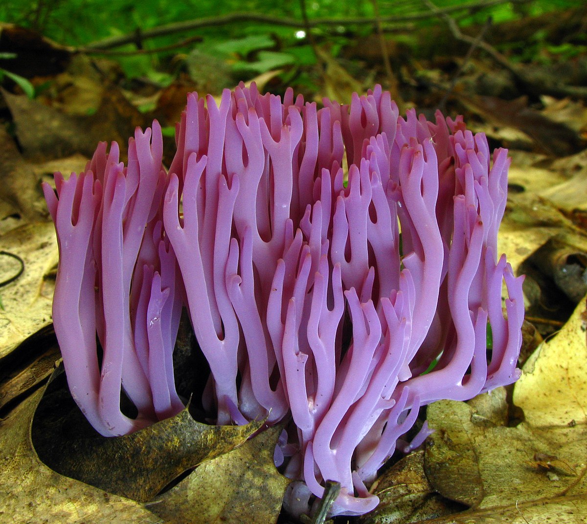


Clavarioid Fungi Wikipedia


1


16 3 Macrofungi Biology Libretexts


Fungus Wikipedia


Club Fungi Reproductive Structures Are Shown In The Image Below What Benefits Are There In Having Brainly Com
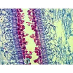


Mushrooms Under The Microscope


The Kingdom Fungi These Morels Are A Type Of Fungus Prized By Many People For Their Distinctive Flavor Unlike The Violets Fungi Are Not Plants And Do Ppt Download
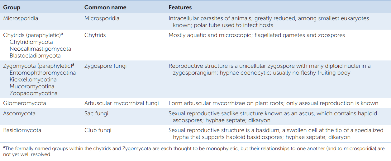


Hillis2e Ch22


Fungus Like Organisms
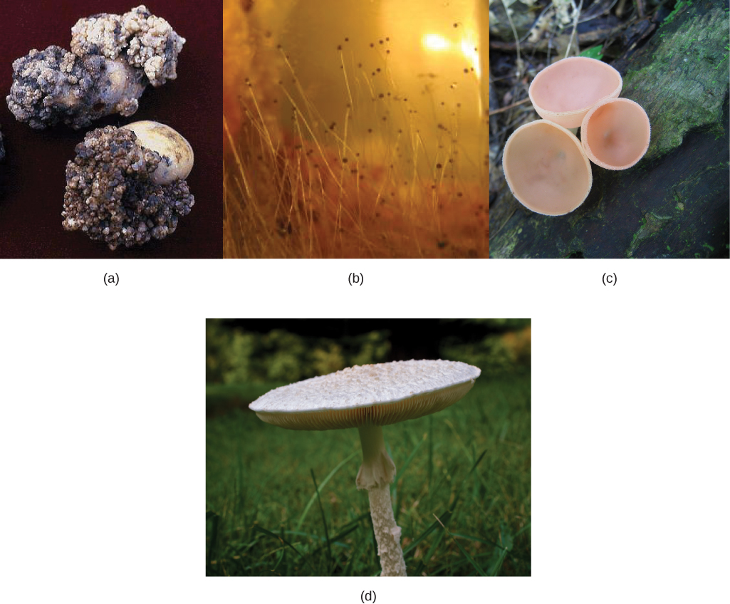


Fungi Concepts Of Biology
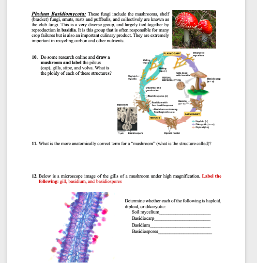


5 What Stage Structure Of Zygomycoya Is Diploid Chegg Com


Basidiomycota


Mu Variant Is Fizzling In Alaska Alaska Public Media


Basidiomycota The Club Fungi


Fungi Antibiotics Yeasts Penicillium


24 3e Deuteromycota The Imperfect Fungi Biology Libretexts


Fungi Basidiomycetes Club Fungi Ctvalley Bio
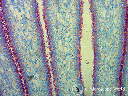


Mushrooms Under The Microscope
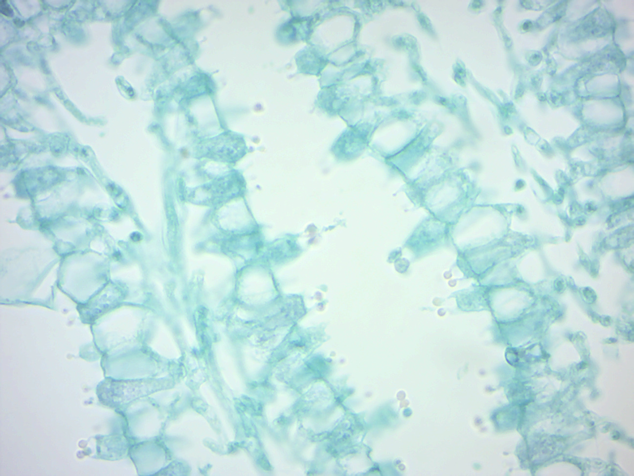


4 Fungi Laboratory Manual For Sci104 Biology Ii At Roxbury Community College


Nick Baker S Hidden Britain Pressreader


Bio 197 Lab 6 Fungi Flashcards Quizlet


Basidiomycetes Club Fungi Coprinus Puccinia Graminis Microscope Slides Carolina Com


Fungi



Fungus Wikipedia


Bio Lab 9 Fungi Microscope Slides Flashcards Quizlet


Reading Fungi Biology Ii Laboratory Manual
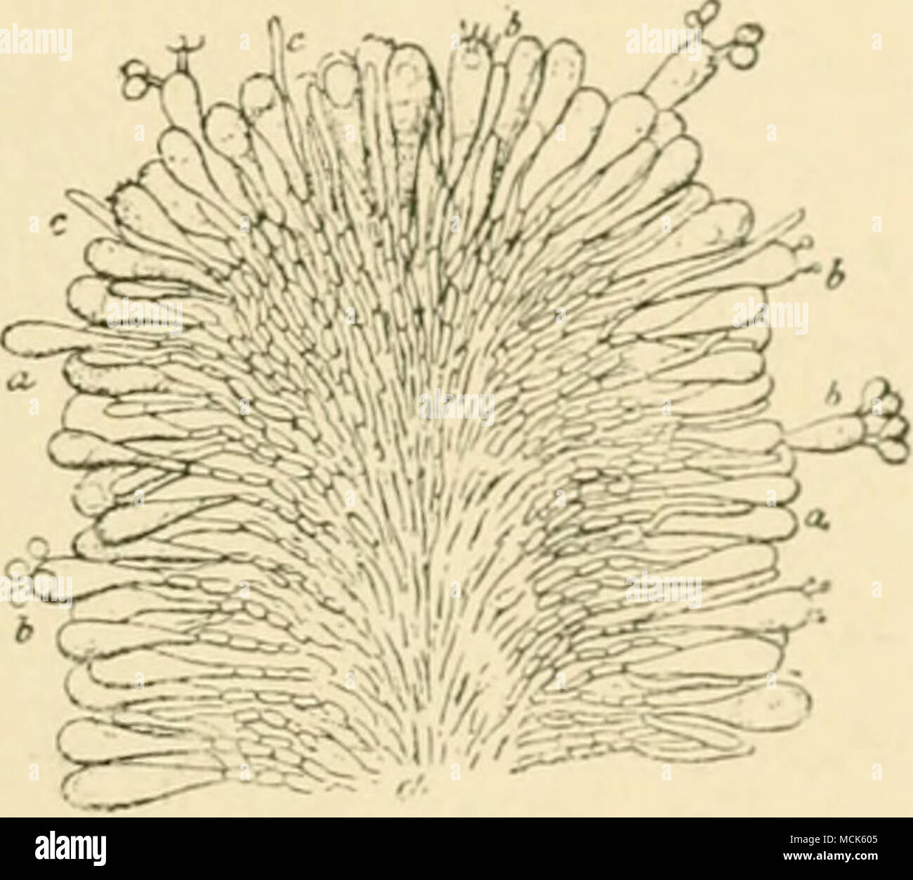


Fig 2s Quot Ar Uricus Meluvx Section Through A Lamella The Hyphae Forming The Subsfcmce Of The Lamella Are Mucli Branched And Send Twigs Outwards Which End In Club Shaped Basidia
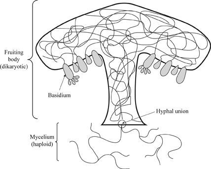


Definition Of The Major Groups Of Fungi Chegg Com


Basidiomycota


Fungi


Club Fungi


Club Fungus Home Facebook
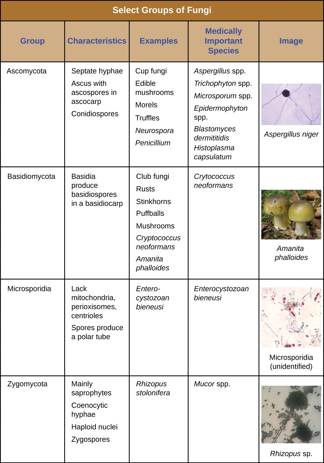


4 3 Fungi Allied Health Microbiology


Basidiomycota
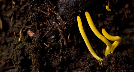


The Secret World Of Fungi The Wildlife Trust For Lancashire Manchester And North Merseyside
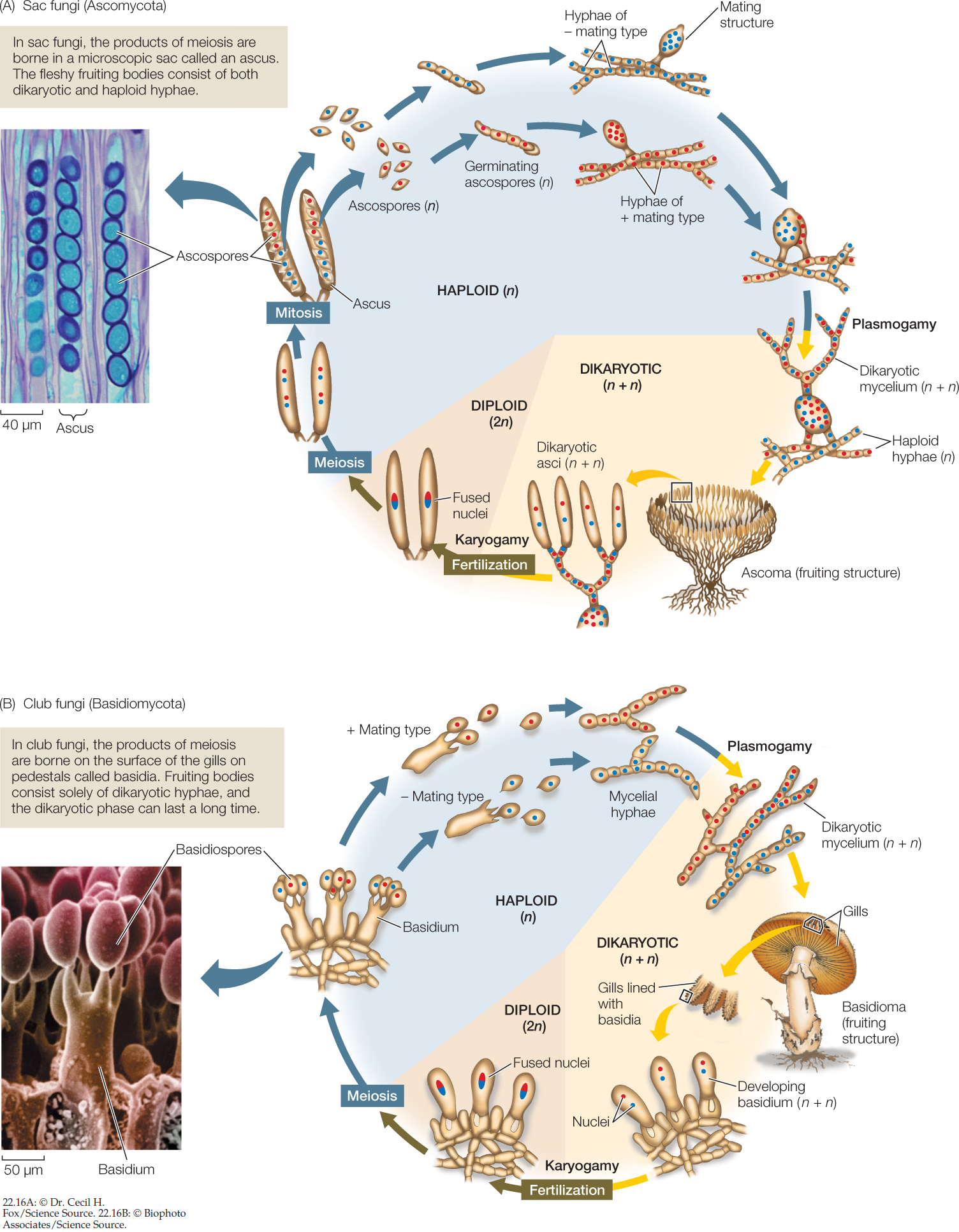


Hillis2e Ch22


Types Of Fungus Classification Of Fungi Phycomycetes Ascomycetes Basidiomycetes Deuteromycetes Neet Youtube


Fungi Basidiomycota The Club Fungi Sparknotes
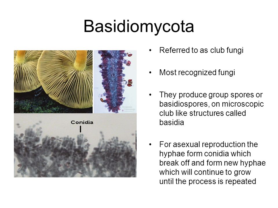


Kingdom Fungi Definition Ppt Video Online Download
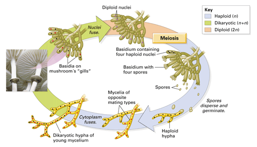


Chapter 18 Concept 18 2


Club Fungi Microscopic Photography Fungi Abstract


Fluorescence Microscopy Journal Club Bruker
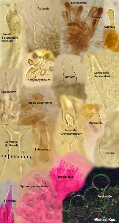


Using A Microscope To Study Mushrooms Mushroomexpert Com
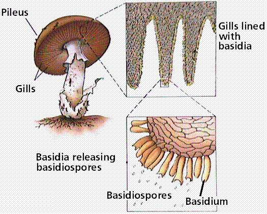


Phylum Basidiomycota The Basidiomycetes Club Fungi


Fungi Basidiomycetes Club Fungi Ctvalley Bio


Bio Lab 9 Fungi Microscope Slides Flashcards Quizlet


Basidiomycota Basidiomycetes Club Fungi Biology 11th Chapter 8 Fungi Youtube
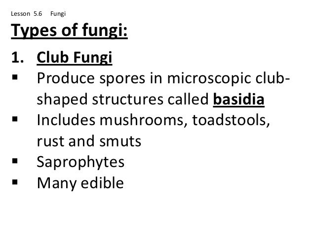


Biology Lesson 5 6


Reading Fungi Biology Ii Laboratory Manual


Pdf Clavariadelphus Pakistanicus Sp Nov A New Club Fungus Basidiomycota Gomphales From Himalayan Moist Temperate Forests Of Pakistan


Fungi Laboratory Notes For Bio 1003


Basidiomycota


Basidiomycetes Club Fungi Instruments Direct
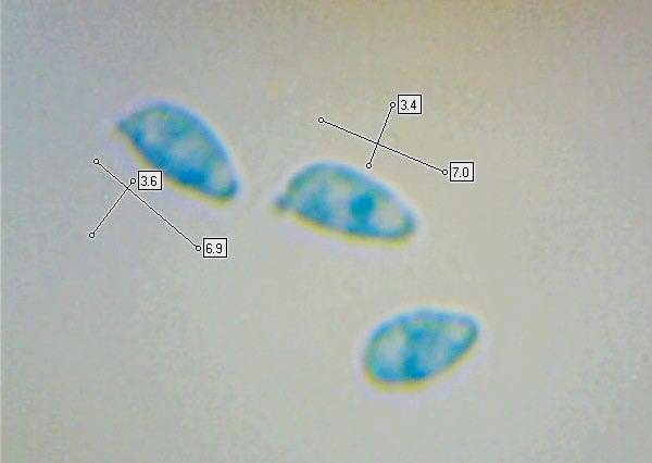


Clavulinopsis Laeticolor Handsome Clubfungus
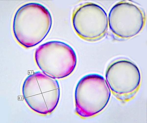


Clavulina Rugosa Wrinkled Club Fungus


Classification And Importance Of Fungi
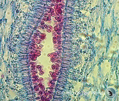


Mushrooms Under The Microscope


Basidiomycota


Www Theexpertta Com Book Files Openstaxbio2e Chapter 24 fungi Pdf


Mushroom Morphology Types Of Identifying Characteristics
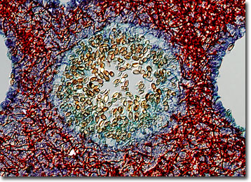


Molecular Expressions Microscopy Primer Specialized Microscopy Techniques Differential Interference Contrast Image Gallery Mushroom Polyporus Fungus
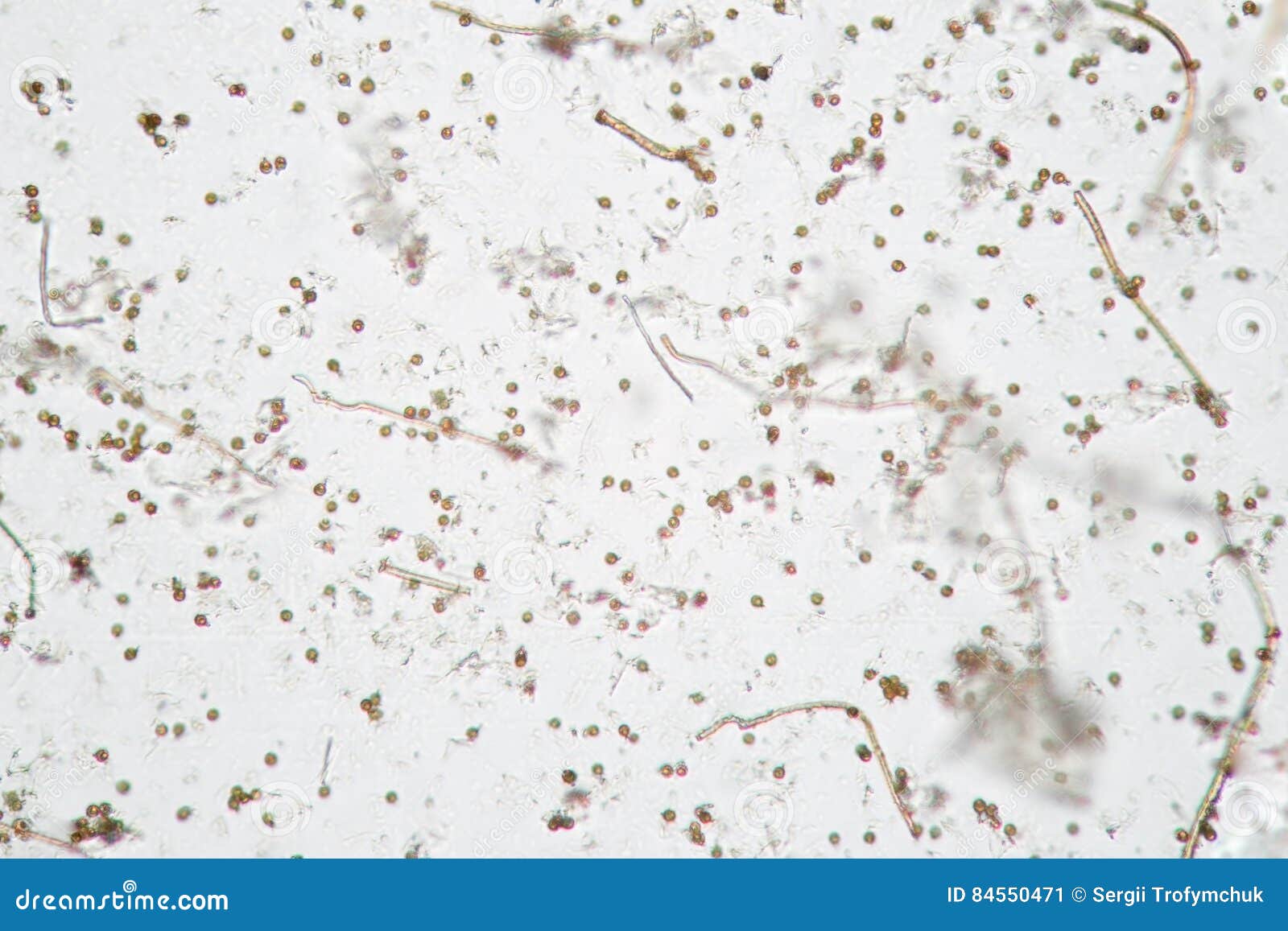


Microscopic Volatile Spores Of Puffball Fungus Lycoperdon Basidiomycota Is A Genus Of Puffball Mushrooms Stock Image Image Of Fungi Particles


Club Fungi Large Images


Experiment 24 General Medical Microbiology Lab


Distinguish Among An Ascus An Ascospore And An Ascocarp An Ascocarp Is The Course Hero


12 Basidium Photos And Premium High Res Pictures Getty Images


Basidiomycetes Club Fungi Instruments Direct
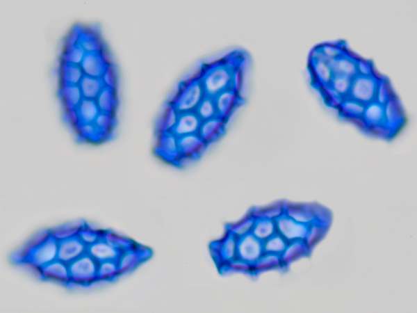


Microscopy Of Spores Hyphae Cystidia Trama To Identify Fungi


The Fungi Kingdom Mycology The Study Of Fungi


Club Fungi Large Images


Fungi Microbiology


Ascomycota Sac Fungi Basidiomycota Club Fungi Victoria Willis


0 件のコメント:
コメントを投稿